


Home » Hospital & Durable Medical Equipment » Ophthalmoscope: Seeing the Light! » Ophthalmoscope: Seeing the Light!
Ophthalmoscope: Seeing the Light!
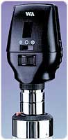
Welch Allyn 3.5 Volt Halogen/Coaxial Ophthalmoscope Head
Retail Price: $701.93
Your Price: $515.54
 Unit: single


Opthalmoscopes enable physicians to examine the interior of the eye. An ophthalmoscope works by directing a tiny beam of light through the pupil, where a physician can then examine the lens, retina, optic nerve, optic disc, blood vessels, and macula for ocular health. Opthalmoscopes can detect abnormalities and pathological changes that signal disease.
The original ophthalmoscope was invented in 1847 by an English mathematician, Charles Babbage. Babbage gave his opthalmoscope to a physician for testing, but it was laid aside and quickly forgotten. Unaware of Babbage’s invention, a German physician-scientist named Hermann von Helmholtz (1821-1894) developed his own version of the ophthalmoscope in 1850. Helmholtz immediately realized and communicated the importance of his new invention and is therefore often credited as the sole inventor. Helmhotz demonstrated the principle of his ophthalmoscope by using a crude device made of cardboard, glue, and microscope glass. By means of this ophthalmoscopes, Helmholtz was able to place the eye of the observer in the path of the rays of light entering and leaving the patient’s eye, allowing the patient’s retina to be seen.
Helmholtz eventually found that looking through the retina into the back of the eye only produced a red reflex. Subsequently, he attached a condenser lens to obtain an inverted image. This image was then magnified five times. He called this mirror and condenser lens combination an indirect ophthalmoscope. It was used regularly for eye examinations until 1920. Helmholtz also invented the ophthalmometer, which was used to measure the curvature of the eye. He also studied color blindness, physiological acoustics, the speed of nervous impulses, and wrote the classic Handbook of Physiological Optics.
Allvar Gullstrand, a Swedish ophthalmologist who studied physiological optics, developed another version of the ophthalmoscope. His innovation was a slit lamp used with a microscope, enabling physicians to locate foreign bodies in the eye.
The modern opthalmoscope is a hand-held instrument containing a small battery-powered lamp that directs a beam of light into the patient’s eye by way of a mirrored prism. The observer looks through a tiny hole in the prism and the instrument, which can be focused by a series of revolving lenses, magnifying the image. The lens focuses the image to give an approximation of the spectacle lenses needed to correct a patient's vision. A recent innovation in this technology is used in eye surgery, whereupon it can project a laser beam to correct a detached retina. Another innovation, called the binocular ophthalmoscope, is used in clinical research, producing images the eye that can be magnified 15 times.
Ophthalmoscopy is invaluable in many fields of medicine, including:
- Cardiology
- Diabetes
- Hematology
- Medical Genetics
- Neurology
- Neurosurgery
- Rheumatology
- Family Medicine
- Pediatrics
- Internal Medicine
- Geriatrics
For patients suffering from chronic headaches, the finding of swollen optic discs, or papilledma, is a key sign that indicates raised intracranial pressure (ICP) which may be due to hydrocephalus, benign intracranial hypertension, or a brain tumor, among other conditions.
For patients with diabetes mellitus, regular ophthalmoscopic eye exams is mandatory to screen for diabetic retinopathy. Vision loss due to diabetes can be prevented by retinal laser treatment if retinopathy is spotted early. In arterial hypertension, hypertensive changes of the retina closely mimic those in the brain, and may predict strokes.