


Home » Hospital & Durable Medical Equipment » Otoscopes: Guiding Light Thru The Ear Canal » Otoscopes: Guiding Light Thru The Ear Canal
Otoscopes: Guiding Light Thru The Ear Canal

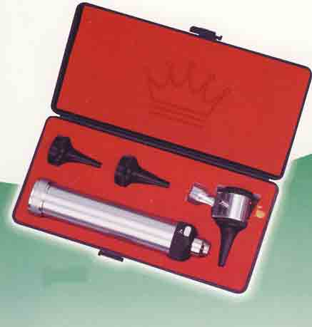
Welch Allyn 3.5V Diagnostic Otoscope Set
Retail Price: $665.47
Your Price: $553.93
 Unit: single


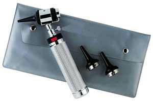
Fiber Optic Mini Otoscope Set
Retail Price: $98.00
Your Price: $73.50
Unit: single
XL 3.5 Uni I Otoscope w/ Charger
Retail Price: $400.65
Your Price: $337.26
Unit: single
Otoscopes are used to examine the ear canal and eardrum. Otoscopes are hand-held instruments that have a tiny light, a magnifying lens, and a funnel-shaped attachment called an otoscope specula. An otoscope consists of three parts:
- Otoscope handles, which contains the power for the light source
- Otoscope heads, which contains the light bulb and magnifying lens
- Specula, which is inserted into the ear canal.
Otoscope exams are normal procedures for most physical examinations. They are also conducted when an ear infection or other such aural problem is suspected due to fever, ear pain, or hearing loss. Otoscopes blow a small puff of air at the eardrum to see if it will respond normally (i.e. vibrate). Otoscopes can also detect a buildup of wax in the ear canal, a rupture of the eardrum, or a puncture in it. Inspection of the eardrum can also provide a lot of information about what is happening within the middle ear - the space within the skull where the hearing and balance mechanisms are situated.
The examination is performed by gently pulling the outer part of the ear upwards and backwards. This straightens the external auditory canal and makes it easier to see the eardrum. A normal external auditory canal has some hair, often lined with yellow to brown wax. The total length of the ear canal in adults is approximately 2cm, giving it a resonance frequency of approximately 3400 Hz, an important frequency region for understanding speech.
The ear canal is normally skin-colored and covered with tiny hairs. The normal eardrum is usually thin, shiny, appears pinkish-gray in color, and is circular in shape. However, an ear infection will cause the eardrum to look red and swollen. In cases where the eardrum has ruptured, there may be fluid draining from the middle ear. A doctor may also see scarring, retraction of the eardrum, or bulging of the eardrum.
Abnormal findings with an otoscope may include:
- A dry, flaky lining suggestive of eczema.
- An inflamed and swollen, narrowed canal, possibly with a discharge indicating infection.
The usual symptoms include itch, local discomfort, a discharge and an unpleasant smell from the ear.
- Qax obscuring the eardrum.
- A foreign body in the ear.
- A perforation in the eardrum.
- Acte infection of the middle ear.
If there is a buildup of wax in the ear canal, it might be rinsed or scraped out with special instruments. However, it is normal for the ear canal to have some yellowish-brown wax. If the Eustachian tube is blocked, air cannot get to the middle ear to equalize the pressure on the eardrum. This can impair hearing or cause pain, if the pressure is enough to stretch the eardrum. If an ear infection is present, the patient may require treatment with antibiotics. The appearance of the eardrum in acute otitis media is dependent on when the infection began. Typically, the eardrum becomes red or yellow in appearance and is opaque with indistinct landmarks. It can also appear to be bulging towards the viewer. Sometimes in acute otitis media the eardrum will burst, reducing the pressure and pain. A tear in the eardrum may be seen as well as a discharge in the outer ear. For the majority of people, such a tear will heal completely.