Home » Hospital & Durable Medical Equipment » Considerations For Brain Anatomy Models » Considerations For Brain Anatomy Models
Considerations For Brain Anatomy Models

Brain Anatomy Model
Retail Price: $273.09
Your Price: $187.47
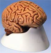
Studying the brain can be very difficult without the use of different types of brain anatomy models. Types or designs of brain anatomy models vary in order to provide the most effective study methods possible, based on the needs of the instructor or the professional. Brain anatomy models, besides being located in science and medical classrooms, may also be found in general and specialized physician's offices as they are helpful in talking to patients about brain trauma, injury and disease.
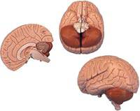
The simplest form of brain anatomy models are the two or four part models. Typically these models are life sized with regards to an adult brain, and they simply divide into the two sides of the brain in the 2 part models or into subsections of the right or left brain in the 4 part models. Another they don't provide as much specific detail they do provide more than sufficient information for introductory medical or science classes plus they are typically all that would be required when discussing the brain with patients. Most of these models are painted so the different brain parts are easy to identify both in the side that comes apart as well as the intact side in the 4 part models. Nerves and major blood vessels in the brain are also highlighted in these models, helping with explanation and medical details for both students and patients.
For more advanced studies there are enhanced models of the particular parts or lobes of the brain. These models typically include larger than life sized but proportional representations of the central brain, which is often impossible to represent in detail in life-sized models to any degree of detail. Many of the specific brain anatomy models that focus on the central brain or the particular aspects of the right or left lobe will be two and a half times life size, providing enough detail for advanced study and understanding. These brain anatomy models are also color coded and painted for easy identification of the various brain parts within the designated area.
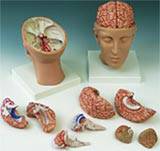
Full heads that also have an options for study of the brain anatomy models are ideal for most teaching situations and even with patient consultations. The models are life size and allow the student or patient to understand the position of the brain parts in relation to the exterior parts of the head. The skull typically lifts off at the top, providing access to the removable parts of the brain. Brain anatomy models of this style also include the major arteries, veins and nerves, highlighted and color coded for easy visualization and understanding.
For neurology students and specialized medical offices there are a wide variety of brain anatomy models that provide highly specialized information. A cerebrospinal fluid circulation model allows the instructor or doctor to discuss the movement of fluid in the brain through the use of a mode. This cross-section model has arrows and color coding to allow easy understanding of how fluid moves through the major parts of the brain, and what possible issues arise if this fluid movement is obstructed or changed.
Finding the correct brain anatomy models for teaching or patient consultation does take some consideration, but they are definitely an asset and an aid to understanding in both situations.
MSEC remains dedicated to providing the very best and the very latest in medical supplies and equipment. We never cease to be on the lookout for the latest innovation that will benefit both our many clients and the patients they dedicate their lives to caring for. If you have any difficulty finding your choices in our vast inventory, call our customer service at 1-877-706-4480 to speed up your order or to make a special request. We are always happy to help you.


 Unit: single


