Home » Hospital & Durable Medical Equipment » Patient Consultations Using a Hip Joint Model » Patient Consultations Using a Hip Joint Model
Patient Consultations Using a Hip Joint Model

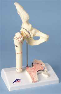
Femoral Fracture & Hip Osteoarthritis Model
Retail Price: $191.85
Your Price: $127.93
 Unit: single

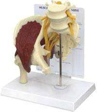
Hip Joint Model w/ Muscles & Sciatic Nerve
Retail Price: $189.33
Your Price: $177.89
 Unit: single
Hip fractures are a very real part of the aging process, especially in Western civilizations. Each year approximately 1,600,000 hip fractures will occur worldwide with 51% of those occurring in Europe and North America. Women are more prone to hip fractures with 75% of the total number worldwide occurring in women over the age of 50. With this high level of occurrence, having the option to clearly show patients what a fracture is essential in any emergency room or patient treatment facility.
Hip fractures can be very difficult for the average person to see on an x-ray or imaging film. It does take an experienced eye to sort out the many bony structures in the pelvis, especially on a flat, black and white X-ray. A better option is to use a hip joint model that clearly allows the patient to see what a normal hip looks like. This also allows the physician to point out, on a 3D model, exactly where the break has occurred. This visual representation will also be instrumental in discussion surgical corrective procedures such as full or partial hip replacements.
There are three different types of hip joint model options that are very useful when discussing issues with patients. The simplest and most streamlined option is the basic hip joint model. This includes the top part of the femur along with the hip joint itself and the corresponding side of the pubis bones. As with most medical anatomical models the right side is typically represented in these 3D models, but custom models may also be possible through specific companies. The simplicity and clarity of this model is perfect for working with patients that need to understand the basic anatomy of the hip joint but may not need additional specific information about the associated musculature.
A slightly more complex hip joint model will include the overlying muscle. It is the same basic model as the simple hip, but the muscles that keep the joint in place are clearly defined on the module. For easy determination and identification the muscles are striated in appearance and are red in color, contrasting sharply with the cream color of the hip bones. This model is ideal for discussing muscle damage, surgical recovery, and muscle regeneration after surgery as well as ligament damage that may occur with some types of trauma.
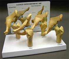
The third type of model is an excellent companion model to the healthy hip joint models mentioned above. It is an arthritic hip model that shows the specific damage and injury that degenerative joint disease, bone spurs, fracture and erosion of the joint can cause to a healthy hip. By comparing the smaller sized diseased or damaged hip joint model miniatures to the healthy hip, the physicians message becomes very clear to the patient. These models are small in size and can easily fit on a desk, counter or on a display case. Using the hip joint model options with the accompanying information cards is a simple way to help your patients understand their diagnosis and why treatment is essential.
MSEC remains dedicated to providing the very best and the very latest in medical supplies and equipment. We never cease to be on the lookout for the latest innovation that will benefit both our many clients and the patients they dedicate their lives to caring for. If you have any difficulty finding your choices in our vast inventory, call our customer service at 1-877-706-4480 to speed up your order or to make a special request. We are always happy to help you.


