Home » Hospital & Durable Medical Equipment » Lungs Anatomy Models Make Teaching A Breath Of Fresh Air » Lungs Anatomy Models Make Teaching A Breath Of Fresh Air
Lungs Anatomy Models Make Teaching A Breath Of Fresh Air

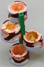
Bronchus Model Showing Asthma & Bronchitis
Retail Price: $104.40
Your Price: $81.40
 Unit: single

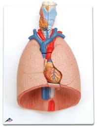
Standard Anatomical Lung Model w/ Larynx
Retail Price: $905.78
Your Price: $744.80
 Unit: single

Chronic Obstructive Pulmonary Disease Model
Call For Pricing 1-877-706-4480
 Unit: single
Anyone that has taught children, young adults or even adult students understands that the more options you have for conveying the information the more learners will absorb, remember and retain. Models are a great visual aid in teaching but they also provide other options for incorporating actual hands on work for the learners in the classroom. The wide variety of lung anatomy models available today can give students of all ages a chance to explore and learn about the lungs and their functioning without having to watch a movie or read about it in a text book.
A lungs anatomy model can range from the very simple versions that basically display healthy lungs on a stand, to specialized models that show the effects of smoking and other respiratory conditions. By having several different types of lungs, anatomy models in the laboratory or classroom allow students to do some comparative work, exhibiting the effects of diseases various health issues.
Lung models that have removable parts are also a great addition to any type of teaching environment. Removable parts allow learners to explore under the surface but still see the lungs in their three dimensional representation, which is often lost when looking at diagrams and charts. Having a better feel and understanding of how major blood vessels, the heart, trachea, larynx, lungs and even the esophagus are placed within the chest cavity can really increase an appreciation for the human body and the systems that are required to work together for healthy functioning. Since the students will see the lungs in actual size reflected by the human lung models as compared to the other organs, a better understanding of the complete anatomy is definitely achieved when these models are part of the standard teaching package.
Segmented lungs anatomy models are perfect for a lot of very detailed types of study for adult learners or those in medical or pre-med classes. These lungs are actually cast from actual human lungs, providing the most lifelike model possible. Each lung, which is attached to the frame and to the bronchial tree, is also divided into 18 different color coded segments. Students can learn about the functioning, position and structure of the smaller parts of the lungs including the bronchioles and alveoli. The segments fit together for secure storage and the stand ensures they can be easily displayed and manipulated, then quickly reassembled for storage.
For a more stylized and less detailed model of the lungs a colorful and highly lifelike standard lungs anatomy model may be just the answer. These durable hard plastic models are perfect for seeing into the lungs and chest cavity with the front halves of the lungs positioned for easy removal. This allows students to look into the chest and remove the larynx, trachea, heart, major veins and arteries as well as the diaphragm. Through being able to work with the model students will definitely have a better understanding of what each organ and blood vessel is in relation to the chest cavity and how the lungs and other systems interact. Adding several anatomical lung models to your classroom or lab is a great way to get students actively involved in learning and allow them to develop a deeper level of understanding.


