Home » Hospital & Durable Medical Equipment » Back Your Discussions Up With A Vertebrae Anatomy Model » Back Your Discussions Up With A Vertebrae Anatomy Model
Back Your Discussions Up With A Vertebrae Anatomy Model

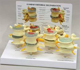
4-Stage Osteoporosis Vertebrae Model
Retail Price: $276.60
Your Price: $221.60
 Unit: 1 set

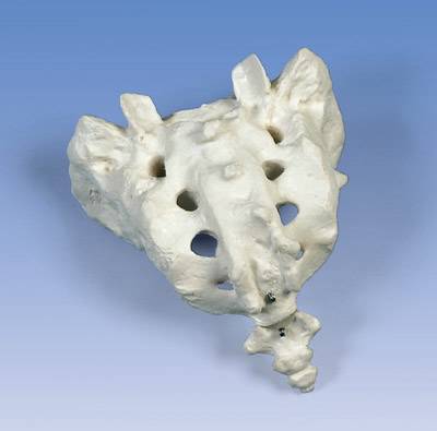
Standard Sacrum and Coccyx Model
Retail Price: $72.20
Your Price: $57.00
 Unit: single
The human spine is made up of many different moving parts, all which have to fit together correctly to allow the human body to move in all directions with ease. When something isn't right in the spine the result is stiffness, loss of range of motion and extreme pain in many cases. Helping patients, students and professionals understand the complex nature of the spine can be difficult at the best of times so using a human vertebrae anatomy model is highly recommended.
Other medical professionals such as physical therapists, sports medicine clinics, rehabilitation therapists, massage therapists and chiropractors may also want to take advantage of having a few different types of vertebrae anatomy models. These models are great to show both a healthy spine and vertebrae as well as what happens with various types of conditions and diseases including trauma to the spinal column.
Depending on the type of medical or classroom environment you work in there are several different options for highly effective models. These can include simple models of two or three different vertebrae in different areas of the spinal column. Often these will show the lumbar vertebrae or the cervical vertebrae. These models, while very specialized, are a great asset in telling students or patients what the issues are that are causing their pain, discomfort or restriction in movement.
As with any model the more options you have for moving, removing and close examination of the various parts of the model the more often you will find the opportunity to use the device. Many models provide the patient and doctor or instructor and student the option to take the model apart. This allows an in depth look into the central area of the spinal column or vertebrae group, including ligaments, muscles, blood vessels, major nerves and the all important discs between the vertebrae. Color coding in realistic yet easy to distinguish colors makes major attributes of the various parts of the spine easy to locate when the model is apart or together.
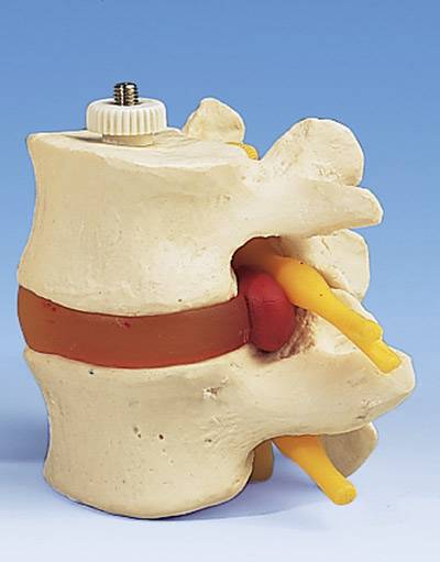
Another interesting option in vertebrae anatomy models is to consider one or more models that also display or clearly depict what injury to the spinal cord looks like. Many of these models have options for showing medical conditions such as vertebral degeneration and disc prolapsed and how those issues cause problems within the spinal cord. The different models may have different discs and vertebras that can be interchanged to highlight how the conditions wear away at the structure of the spine and result in long term permanent damage.
Some of the vertebrae anatomy model kits on the market are very realistic in appearance. As opposed to the more colorful and traditional model types these kits strive to be a realistic as possible, generally designed to show the vertebrae themselves rather than the surrounding soft tissues. These models are typically one a simple stand or on a molded base that provides easy storage as well as easy access to each vertebrae as needed for explanation or study.


