Home » Hospital & Durable Medical Equipment » Human Eye Models For Teaching Patients Or Students » Human Eye Models For Teaching Patients Or Students
Human Eye Models For Teaching Patients Or Students
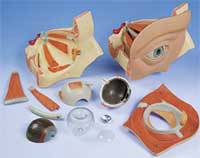
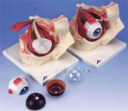
The human eye is one of the most complex systems in the body, with each of the many layers and tissues of the eyeball all required to produce the nerve impulses that the brain decodes into what we see. The human eye models can be manipulated and students may interact in a way that makes the learning process fun and informative.
There are many different options when choosing human eye models for any type of classroom, patient care room, or lab setting. An ideal situation is to have several different types of models which could be used as needed, rather than having to rely on just one. Basic information and understanding could be easily conveyed using a simple model with just a few parts and sections while a more complex medical issue or anatomical class would definitely benefit from a multi-section model.
A seven or eight part human eye model provides a lot of detail for patients and students to examine all the various internal and external features of the eye and how they interact within the eyeball itself. In most models the removable options will include the lens, choroid, iris, retina, sclera, cornea, eye muscle attachments and the vitreous humour. The real benefit of these eye models is that they can be three or five times life size, allowing easy vision into the smallest details of the eye. Some models also include the tear system, which can be a significant feature in teaching and explaining problems to patients.
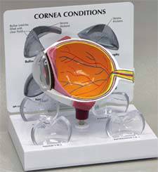
While the above mentioned human eye models are designed to be lifelike, there are also different types of models that show how the eye functions in focusing and in providing corrective lenses or other medical procedures. A functional human eye model is designed so that the lens and ciliary body can actually change shape, allowing students and patients to visually see how many common vision problems occur. This includes near and far sightedness and even how aging affects the lens's ability to focus, resulting in poor vision.
We carry eye models with the ability to project pictures on the retina. The malfunctioning of the lens in providing focus is easy to see even for someone without a medical background. Artificial lenses, which represent corrective glasses or contacts, can then be inserted into a stand in front of the retina. The patient or student will easily be able to visualize how the glasses will help in improving vision.
For very specialized types of eye diseases and conditions a micro-anatomical eye model is a great option. These are typically used in training classrooms and in specialists' offices, but they do provide the detail in the retina that clearly shows the internal structure at a microscopic level. Since these concepts are often very difficult for a patient or student to understand, the model helps to provide the necessary details that simply would be to difficult to grasp without the visual aid.





 Unit: single

